


SpectraSplit® 7
SpectraSplit® 7 are patented filter sets that empower fluorescence microscopes with seven color channels. The channels are distributed over four filter sets and specifically separate the most commonly used fluorochromes in immunofluorescence (see table below). Hence, you can immunostain tissues with up to seven fluorochromes and still visualize each fluorochrome in the sample with SpectraSplit® 7 without bleedthrough. No additional software and no computational spectral unmixing are required.
SpectraSplit® 7 can easily be installed in both standard and scanning fluorescence microscopes, and can be run with different popular light sources. See the description below for more information.
Cat no. 7001991-1
Request a quote or information
Request a quote or order
Request a quote or order with payment by invoice by sending us an email via the button above. When ordering, please include information on the products you wish to order, organization, VAT number (only EU countries), delivery address, and invoice address. Please also state contact person and phone number for delivery by courier.
We ship globally – a shipping cost of €50 is added to countries within the EU and €80 to all other countries. We can also charge in $USD.
NB: VAT is not added to the final cost under Article 138 VAT directive (other EU countries) and 146 VAT directive (export to non-EU countries).
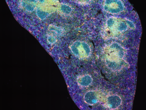
The SpectraSplit® 7 filter sets are highly efficient in separating the emission signals of seven different fluorochrome classes, with spillover between channels being less than 0.5%. As a result, high-contrast images are generated without the need for spectral unmixing or any other bleed-through corrections.
SpectraSplit® 7 immediately empowers your microscope with seven independent color channels that each generates crystal clear images. Importantly, you do not need to be a fluorochrome expert or microscope guru to produce 7-color images.
Fluorochromes
SpectraSplit® 7 is fully compatible with commonly used fluorochromes, such as AlexaFluor® dyes, as well as the classic FITC/Cy3/Cy5/Cy7 dyes and many of the OpalTM dyes.
In addition, our StreptaClick® reagents for multiplex IHC have been optimized to be compatible with SpectraSplit® 7 for easy 7-color assay development to make the most of your microscope or scanner equipped with SpectraSplit® 7.
Fluorochromes compatible with SpectraSplit® 7
| Channel | Traditional dyes | StreptaClick® dyes | ThermoFisher | Biotium | ThermoFisher | Atto-tec | Akoya | Ultivue | Sirigen/BD | Protein expression fluorophores |
| S-Split Blue (375) | DAPI | DAPI, Tyramide 405 | Alexa Fluor 405 | CF405 | Dylight 405 | DAPI | DAPI | BV421 | Hoecht | |
| S-Split Cyan (435) | CFP | Tyramide CF®430 | CF430 | Atto425 | Opal p.480 | BV480 | cCFP | |||
| S-Split Green (490) | FITC | Tyramide 488, Color 488 | Alexa Fluor 488 | CF488 | DyLight 488 | Atto488 | Opal 520 | Ultivue, FITC | eGFP | |
| S-Split Orange (545) | Cy3/TRITC | Tyramide 555, Color Atto 542 | Alexa Fluor 546 | CF555 | DyLight 549 | Atto542 | Opal 570 | Ultivue, Cy3 | mOrange/ mRFP |
|
| S-Split Red (590) | Texas Red | Tyramide 594, Color 594 | Alexa Fluor 594 | CF594 | DyLight 594 | Atto590 | Opal 620 | mCherry/mRaspberry /mPlum |
||
| S-Split Far-red (650) | Cy5/Cy5.5 | Tyramide 647, Color 647 | Alexa Fluor 647 | CF647/680 | DyLight 649 | Atto647/665 | Opal 690 | Ultivue, Cy5 | miRFP703 | |
| S-Split Infra-red (740) | Cy7 | Styramide iFluor 750 | Alexa Fluor 750 | CF750 | DyLight 750 | Atto740 | Opal p.780 | Ultivue, Cy7 |
*Cy3, Cy5, Cy7 are registered trademarks of GE Healthcare, Alexa Fluor and DyLight dyes are trademarks of Thermo Fisher Scientific, CF dyes are trademarks of Biotium, Atto dyes are trademarks of Atto-tec, Opal dyes are trademarks of Akoya Biosciences, Brillialt Violet dyes are trademarks of Sirigen/BD

Bandwidth
The approximate mid excitations for the different channels are at 375, 435, 490, 545, 590, 650, and 740nm. However, the exact bandwidths are proprietary information and not publicly disclosed.
Microscopes and light sources
SpectraSplit® 7 is designed to work with both standard and scanning fluorescence microscopes and can be found in scanners from multiple manufacturers including Evident (VS200), TissueGnostics (TissueFAXS CHROMA), and Nikon (Ti2). It is compatible with various lighting sources including pE-800 and pE-4000 from CoolLED, X-Cite NOVEM from Excelitas, and the new SPECTRA X Light Engine from Lumencor.
Hardware
SpectraSplit® 7 contains four custom made filter sets, manufactured to the high product standards of Chroma Technologies. SpectraSplit® 7 is a patented technology, exclusive to our brand. These filter sets are not available under any other name. Each filter set includes one excitation filter, one emission filter (25 mm in diameter), and one dichroic mirror (25.5 x 36.0 x 1 mm).
Set 1: Triple-band set (375/490/740)
Set 2: Double-band set (435/650)
Set 3: Single-band set (545)
Set 4: Single-band set (590)
Ordering information
| Cat no. | Product |
| 7001991-1 | SpectraSplit® 7 |
Please contact us to inquire about custom filter configurations.
RESOURCES
FOLDER
SpectraSplit® 7 folder
PUBLICATIONS
2026
2025
2024
2023
2022
2021
2019
2018
2017
** The original publication demonstrating what is now our SpectraSplit® bandpass filters for multiplex immunofluorescence microscopy.
Is your publication utilizing a Kromnigon product not listed on our site? Please let us know!
Related products

BiotinPure™ Antibody Biotinylation Kit
The BiotinPure™ Antibody Biotinylation Kit is a complete kit for easy biotinylation of antibodies at room temperature within 1 hour. The kit uses amine/ester chemistry to attach 2-3 biotins per antibody and includes a purification wash step that removes unreacted free biotin from the final biotinylated antibody solution.
The antibodies to be biotinylated should be free from other proteins, such as BSA.
The BiotinPure™ Antibody Biotinylation Kit is designed for use with StreptaClick® reagents. The biotinylated antibodies may also be used for other applications.
Cat. no. 9000051-1
Order or request further information
Request a quote or order
Request a quote or order with payment by invoice by sending us an email via the button above. When ordering, please include information on the products you wish to order, organization, VAT number (only EU countries), delivery address, and invoice address. Please also state contact person and phone number for delivery by courier.
We ship globally - a shipping cost of €50 is added to countries within the EU and €80 to all other countries. Upon request we can also charge in $USD for payments by invoice.
Webshop
Orders can also be placed through our webshop via the form below. Once a product has been added a popup will appear with the choice of proceeding to checkout or adding additional products. The shopping cart icon at the top right corner also takes you to checkout.
At checkout you will have the choice of secure payment by credit card via Stripe, Apple Pay, Google Pay or invoice. Shipping charges are added automatically.
NB: VAT is not added to the final cost under Article 138 VAT directive (other EU countries) and 146 VAT directive (export to non-EU countries).
Select your BiotinPure™ Kit:

SpectraSplit® 10 filter sets
SpectraSplit® 10, developed from our patented SpectraSplit® 7 filter sets, will revolutionize fluorescence microscopy as 10 channels can be directly imaged without spectral unmixing. The channels are distributed over five filter sets and separate the most commonly used fluorochromes in immunofluorescence, in addition to channels anticipated to gain popularity in the near future (see table below). Tissues and other sample types can be immunostained with up to 10 fluorochromes, visualized without bleedthrough. No additional software and no computational spectral unmixing are required.
SpectraSplit® 10 can easily be installed in both standard and scanning fluorescence microscopes.
Request a quote or information

StreptaClick® 8-plex for ZEISS Axioscan 7 spatial biology
The StreptaClick® 8-Plex Multiplex IHC Kit provides a rapid and heat-treatment-free Tyramide Signal Amplification (TSA) staining cyclic workflow for frozen and FFPE tissue sections. It is optimized for imaging on the ZEISS Axioscan 7 spatial biology platform, enabling visualization of up to eight colors in a single run. The kit is ideal for researchers in high-throughput laboratories who require a reliable, time-efficient, and flexible multiplex immunohistochemistry workflow.


StreptaClick® Color
StreptaClick® Color is a modified streptavidin that labels biotinylated antibodies with fluorochromes in solution, mimicking directly conjugated antibodies as we have removed the issue of antibody aggregation through monovalent binding. StreptaClick® Color provides a rapid, robust, and flexible method for attaching fluorochromes to biotinylated antibodies. Once labeled with StreptaClick®, the antibody is stable and can be used in the same manner as antibodies that are directly conjugated with fluorochromes. The labeling reaction is highly efficient and can be carried out at any volume, concentration, or buffer.
Order or request further information
Request a quote or order
Request a quote or order with payment by invoice by sending us an email via the button above. When ordering, please include information on the products you wish to order, organization, VAT number (only EU countries), delivery address, and invoice address. Please also state contact person and phone number for delivery by courier.
We ship globally – a shipping cost of €50 is added to countries within the EU and €80 to all other countries. Upon request we can also charge in $USD for payments by invoice.
Webshop
Orders can also be placed through our webshop via the form below. Once a product has been added a popup will appear with the choice of proceeding to checkout or adding additional products. The shopping cart icon at the top right corner also takes you to checkout.
At checkout you will have the choice of secure payment by credit card via Stripe, Apple Pay, Google Pay or invoice. Shipping charges are added automatically.
NB: VAT is not added to the final cost under Article 138 VAT directive (other EU countries) and 146 VAT directive (export to non-EU countries).
Select your StreptaClick® Color Kit:


StreptaClick® HRP
StreptaClick® HRP Multiplex IHC Kit is an accelerated heat-treatment-free multiplex Tyramide Signal Amplification (TSA) staining solution for up to nine markers (8 antibodies + DAPI) for fixed cells, FFPE samples and frozen tissues. This comprehensive kit is designed for researchers who demand ease of use and flexibility in multiplex immunohistochemistry (IHC).
The kit includes everything needed to label biotinylated primary antibodies with horseradish peroxidase (HRP) enzyme in solution utilizing our modified streptavidin StreptaClick®. As StreptaClick® removes the issue of antibody aggregation through monovalent biotin binding it is particularly suited for labeling primary biotinylated antibodies. The kit also features a novel HRP Quench Buffer that eliminates the need for antibody-stripping by heat-treatment such as boiling between cycles. This feature maintains tissue morphology over multiple staining cycles making it ideal for both frozen and FFPE tissue sections, and higher degrees of multiplexing than comparable solutions.
In combination, these features makes the protocol highly automation-friendly, while results for manual staining of six markers + DAPI are achievable in one day.
Order or request further information
Request a quote or order
Request a quote or order with payment by invoice by sending us an email via the button above. When ordering, please include information on the products you wish to order, organization, VAT number (only EU countries), delivery address, and invoice address. Please also state contact person and phone number for delivery by courier.
We ship globally – a shipping cost of €50 is added to countries within the EU and €80 to all other countries. Upon request we can also charge in $USD for payments by invoice.
Webshop
Orders can also be placed through our webshop via the form below. Once a product has been added a popup will appear with the choice of proceeding to checkout or adding additional products. The shopping cart icon at the top right corner also takes you to checkout.
At checkout you will have the choice of secure payment by credit card via Stripe, Apple Pay, Google Pay or invoice. Shipping charges are added automatically.
NB: VAT is not added to the final cost under Article 138 VAT directive (other EU countries) and 146 VAT directive (export to non-EU countries).
Select your StreptaClick® HRP Kit:


StreptaClick® Precision
StreptaClick® Precision is a modified streptavidin designed for precise stoichiometric 1:1 conjugations. Currently, StreptaClick® Precision is available with exactly one azide or exactly one DBCO (dibenzylcyclooctyne). StreptaClick® Precision also features a unique cis-divalent design for enhanced monovalent binding to biotinylated proteins. In addition, the conjugation site and the biotin binding region are on opposite sides of StreptaClick® Precision, minimizing the risk of conjugates interfering with biotin binding. This design makes StreptaClick® Precision ideal for attaching bulky molecules without sterically hindering biotin binding and without causing aggregation when mixed with biotinylated antibodies in solution.
Click-chemistry ready
StreptaClick® Precision is currently available with a single azide or DBCO group for either copper(I)-catalyzed azide-alkyne cycloaddition (CuAAC) or copper-free strain-promoted azide-alkyne cycloaddition (SPAAC) click chemistry.
For oligonucleotides, we strongly recommend SPAAC click chemistry (DBCO/azide).
Pre-conjugated, off-the-shelf
StreptaClick® Precision based, pre-conjugated products are also available. These include StreptaClick® Precision with a single 14 mer DNA oligonucleotide or a single bright fluorochrome (488 nm or 647 nm).
Custom conjugation as a service
Please inquire to discuss the feasability of your project.
Order or request further information
Request a quote or order
Request a quote or order with payment by invoice by sending us an email via the button above. When ordering, please include information on the products you wish to order, organization, VAT number (only EU countries), delivery address, and invoice address. Please also state contact person and phone number for delivery by courier.
We ship globally – a shipping cost of €50 is added to countries within the EU and €80 to all other countries. Upon request we can also charge in $USD for payments by invoice.
Webshop
Orders can also be placed through our webshop via the form below. Once a product has been added a popup will appear with the choice of proceeding to checkout or adding additional products. The shopping cart icon at the top right corner also takes you to checkout.
At checkout you will have the choice of secure payment by credit card via Stripe, Apple Pay, Google Pay or invoice. Shipping charges are added automatically.
NB: VAT is not added to the final cost under Article 138 VAT directive (other EU countries) and 146 VAT directive (export to non-EU countries).
Select your StreptaClick® Precision Kit: